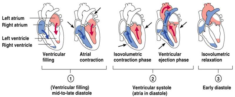
Contraction and Relaxation of Cardiac Fibre
Contraction of the cardiac fibers occurs through the sliding filament model of contraction. Some of the initial steps in the contraction and relaxation reaction include the excitation, as well as the shortening of muscular fibers. During contraction process, significant elements include the thick filaments of the contractile protein myosin and thin filaments of actin. Further, troponin binds to the tropomyosin hence forming a functional unit called troponin-tropomyosin which participates as a regulatory protein. In the process of diastole, the troponin-tropomyosin is attracted to act in hence preventing the interaction between myosin and actin. Notably, the surface of troponin possesses a receptor that binds to calcium. On the other hand, when the receptor in not occupied with calcium ion, it prohibits actin and myosin interaction. When action potential proceeds the systole cycle, it increases the concentration of calcium in the cytoplasm. Furthermore, calcium is bound with troponin hence allowing troponin-myosin complex to get detached from actin. As a result, the molecules of myosin are supported thus shifting act in and myosin filaments to move towards opposite directions.
Calcium is the contraction inducer of the cardiac fibers. It originates from sources such as extracellular space of sarcolemma-glycocalyx and sarcoplasmic reticulum formed by a system of vesicles. Also, adenosine triphosphate plays an important role during contraction as well as relaxation processes. When the cardiac fibers are relaxed (diastole), the adenosine triphosphate gets bound to the myosin molecule hence releasing troponin-myosin complex. Consequently, the process of relaxation is achieved through two mechanisms. Nevertheless, the concentration of calcium levels in the cytoplasm affects the contractile protein in various ways; they raise inhibitory effect of troponin-tropomyosin on actin. Besides, calcium ions activate the enzyme myosin ATP-ase, which is present in the myosin molecule. It helps in the process of splitting ATP molecule to the lesser Adenosine diphosphate. In most cases, the process may suppress the formation of the actin-myosin. At the same time, the energy from the adenosine molecule is acquired for contraction. Thus, sliding of the thin filaments between the thick filaments of myosin occurs. On the other hand, diastole occurs when the filaments return to the previous position.
To sum up, the filaments of actin as well as myosin proteins are the ones responsible for the process of contraction. When the cardiac fibers relax, two systems inhibit the smooth interaction of between the proteins. Although the troponin-tropomyosin complex binds to the actin, the adenosine triphosphate is attracted to the contractile protein myosin. Similarly, calcium concentrations in the cells bind with troponin that activates the myosin ATP-ase enzyme to split Adenosine triphosphate molecule. Therefore, smooth interaction between actin and myosin is maintained. Cardiac fibers regulate the flow of blood as well as oxygen throughout the body.

