Effect of various B-mode controls to optimize the ultrasound images
The behavior of sound is what makes imaging possible. Sound moves in waves which can bounce back a term known as echo when they hit an object. The type of bouncing back or echo that is realized from a specific sound wave is what will be used to know the features of the object it bounced back from. This principle is applied when a transducer is used in a medical examination. While moving the sound waves will meet obstacles like body organs and tissues and bounce off them. The bouncing off produces echoes which are picked up a microphone which records the type of echoes sent and transmits the information to a computer for further analysis. (Robertson, 2001).
Optimization of the images produced by ultrasound causes clearer analysis of the degree of contrast and clarity of the image produced. This causes the image to have a higher resolution enabling better analysis of it on the grounds shape and size to be used for diagnosis (Joseph, 2002).
Sonography modes and B-mode controls
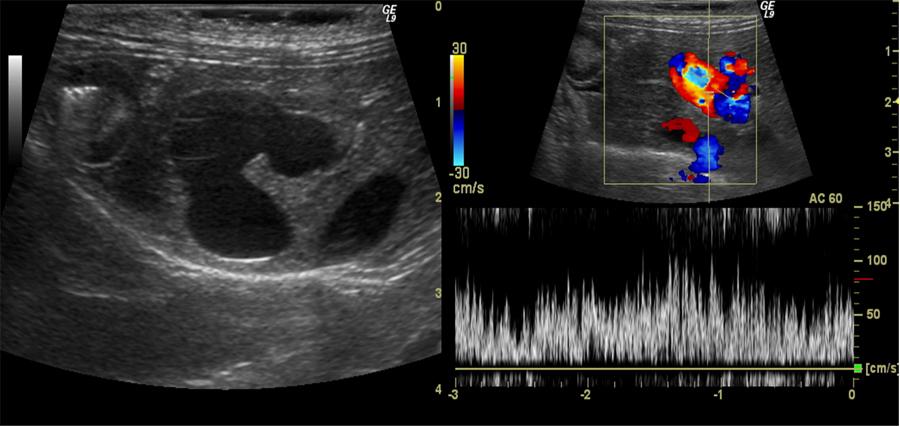
Medical imaging employs several techniques or sonography modes. These modes are determined by the movement of the transducers specifically piezoelectric transducers used in medical imaging. The transducers of either A mode, B Mode, C Mode, M Mode, Pulse inversion mode or Harmonic mode all employ a similar property in the movement of the sound waves. In all the modes, the method with which a signal was sent is the same method with which the signal bounces or echoes back to bring the desired information (Joseph, 2002). B –Mode can utilize color maps to increase the contrast range and levels which in turn optimizes the imaging (Charles, Beckmann, Rodger, &, Barbara, 2009). The imaging that involves color production boosts up the general contrast levels; this in turn improves on the appearance of the organs and tissues being visualized unlike the previous use of grey images (American college of Radiology, 2011).
The image produced by ultrasound can be improved by focusing on the basic aspects and principles of the transducer used. These aspects that constitute the settings of B-mode, by being manipulated in various angles as discussed below will bring out an image with a higher contrast and resolution level. The idea will be to optimize resolution and signal strength which will produce an optimal image (Baker, 2007; Robertson, 2001).
Acoustic Power
Synthetic aperture is an idea that can be utilized with minimal percentage of errors in creating ultrasound images. This line of thought makes the ultrasound system simpler and cheaper. Acoustic power is useful since ultrasound waves display properties similar to those of conventional waves. Sound waves being longitudinal waves cause acoustic power to have its particles displaced in the direction of travel of the waves. These waves also tend to display properties similar to those of light incident ultrasound waves through a body tissue resulting in a refracted ultrasound wave being transmitted to the other plane with an altered line of progression known as reflected ultrasound (American college of Radiology, 2011)..
The effects repeats itself over various tissues with changing from one tissue interphases to another, and the consequently changes the properties of an ultrasound wave known as acoustic properties (Baker, 2007; Robertson, 2001). The travelling energy or properties of a sound wave is reduced as the ultra sound wave bounces off different material with different elemental properties. This quality can be used to optimize the quality of an ultrasound image by controlling the tissues it has to pass through before reaching the targeted area (Baker, 2007; Robertson, 2001).
Frequency
Increasing the frequency of the transducer will produce clearer and sharp images. This will however cause a problem because when frequency is increased the ability of the sound wave to reach to structures located deeper in the body is hampered hence only visualization of superficial structures. It should be noted that frequency is determined by the highest amount of time taken from the source to the target (John, Jan, Maurice, & Yona, 2007).
Focal Points
Focusing the ultrasound wave can also increase the clarity of the image produced. This can be done at a given distance that is exact from the equipment being used in this case a transducer. All the tissues and objects located within the focal point will eventually produce more clear images (Carolyn, Dennis, Ronald, 2005).
Penetration
Determining the number of pulse produced per second will give one an idea of the penetration of a pulse which determines the penetration of a given emitted wave. Transducers have to first of all record all the images from a given pulse before sending out another pulse in order to avoid confusion by having so many images returning at the same time (Robertson, 2001). So in every transmission, penetration detector, RPF determines the images by quantity and stores in the memory. This causes the RPF to vary directly with the penetration rate and adjusting them to their optimal will increase the quality of the ultrasound images generated (Bricker, 2000).
Other qualities of transducer that can be regulated to come up with a clearer image include angle sector width which controls the size of the section to be swept with a wave. Another quality is altering the tilt, the gain and the time gain compensation. The above will enhance view of structures lying on the sides, increase amplification for the electrical pulse and cause a differentiation of gain adjustments to take care of variation in time taken to travel through the structures respectively. The power output of the machinery used can also be used to optimize image production.
